Fluorescence Resonance Energy Transfer (FRET) Application Guide
Basics of FRET
Fluorescence resonance energy transfer (FRET), also known as Förster resonance energy transfer, is a phenomenon whereby an “excited” donor fluorophore molecule transfers energy in a non-radiative process to a suitable acceptor fluorophore molecule. This transfer of energy is termed “non-radiative” because it occurs without the emission of a photon from the donor fluorophore. Instead, it involves the direct transfer of energy of a donor molecule in the excited state when its fluorescence emission spectrum overlaps with the excitation spectrum of an acceptor fluorophore. The resulting FRET signal from the acceptor is often referred to as “sensitized emission”.
FRET occurs between donor and acceptor molecules only after several conditions are met. First, the molecules must be sufficiently close – typically <10 nm, but more often a closer apposition is required to successfully demonstrate FRET. This distance dependence makes FRET a powerful tool, as such close apposition between molecules is often evidence of molecular interaction. A FRET signal is direct empirical evidence of this close physical relationship, independent of the resolution of the imaging system and independent of conventional colocalization techniques. The Förster distance is the distance at which the efficiency of energy transfer is 50% – typically between 4-7 nm, depending on the particular donor/acceptor pair, and is typically used to characterize the potential usefulness of a fluorophore pair. The efficiency of energy transfer increases exponentially as molecules become closer, and is exquisitely sensitive to the distance of separation.
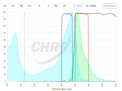
Second, as mentioned above, the excitation spectrum of the acceptor must overlap with the emission spectrum of the donor. Greater spectral overlap generally increases the likelihood of FRET, but also complicates data analysis by increasing the risk of false-positive signals due to direct excitation of the acceptor.
Third, the donor and acceptor molecule dipoles must be oriented favorably in order for FRET to occur. In addition to proximity, the dipoles must be favorably aligned for efficient energy transfer. Most often this condition is met randomly with large populations of molecules and a standard value for dipole orientation is used in most FRET calculations.
These requirements make FRET particularly sensitive to spectral overlap and signal separation, placing strict demands on filter selection.
Total internal reflection fluorescence (TIRF) microscopy places unusually high demands on emission filters and dichroic mirrors. Unlike standard widefield setups, the reflected excitation beam travels back through the objective — meaning emission filters must block excitation energy they'd never see in other configurations, and dichroic mirrors must maintain flatness specifications that are difficult to achieve with standard manufacturing methods.
Filter Requirements for FRET
Excitation Filters
FRET is an imaging technique which can benefit greatly by the appropriate choice of filters. In widefield imaging applications, donor excitation filters should be chosen to avoid direct excitation of the acceptor molecule as much as possible. While it’s never possible to entirely avoid direct excitation of an acceptor fluorophore, carefully chosen exciters will best exploit the difference between the excitation spectra of the donor and acceptor.
For applications involving laser excitation, such as confocal and TIRF imaging, the most appropriate laser wavelengths should be used with appropriate laser clean-up filters.
Emission Filters
Likewise, emission filters should be chosen to best separate the donor /acceptor emission spectra.
For example, in some cases it may be best to red-shift the acceptor bandpass to minimize the detection of donor fluorescence emission, while in other cases this will limit detection of the acceptor emission too much and will be counter-productive.
*Note about FRET Microscopy
FRET is a computation-intensive technique involving much image processing and image correction. It is not a simple technique by any means, and is best performed using systems with external filter wheels which afford independent control of excitation and emission wavelengths, or image-splitting systems where donor and FRET signals may be acquired simultaneously in live cell imaging. Temporal resolution may be critical in FRET, depending on the dynamics of the model system. Both approaches will require the use of polychroic mirrors which reflect multiple excitation bands and transmit multiple emission bands.
Additional techniques such as multiphoton excitation microscopy and fluorescence lifetime imaging microscopy (FLIM) are also used in FRET applications, each with their own advantages and disadvantages.
Additional Resources

Still Have Questions?
Our applications team works with FRET setups across most major commercial platforms. Tell us your instrument and fluorochromes and we'll point you in the right direction.